

X-ray showing severe arthritis of the ankle joint (front)
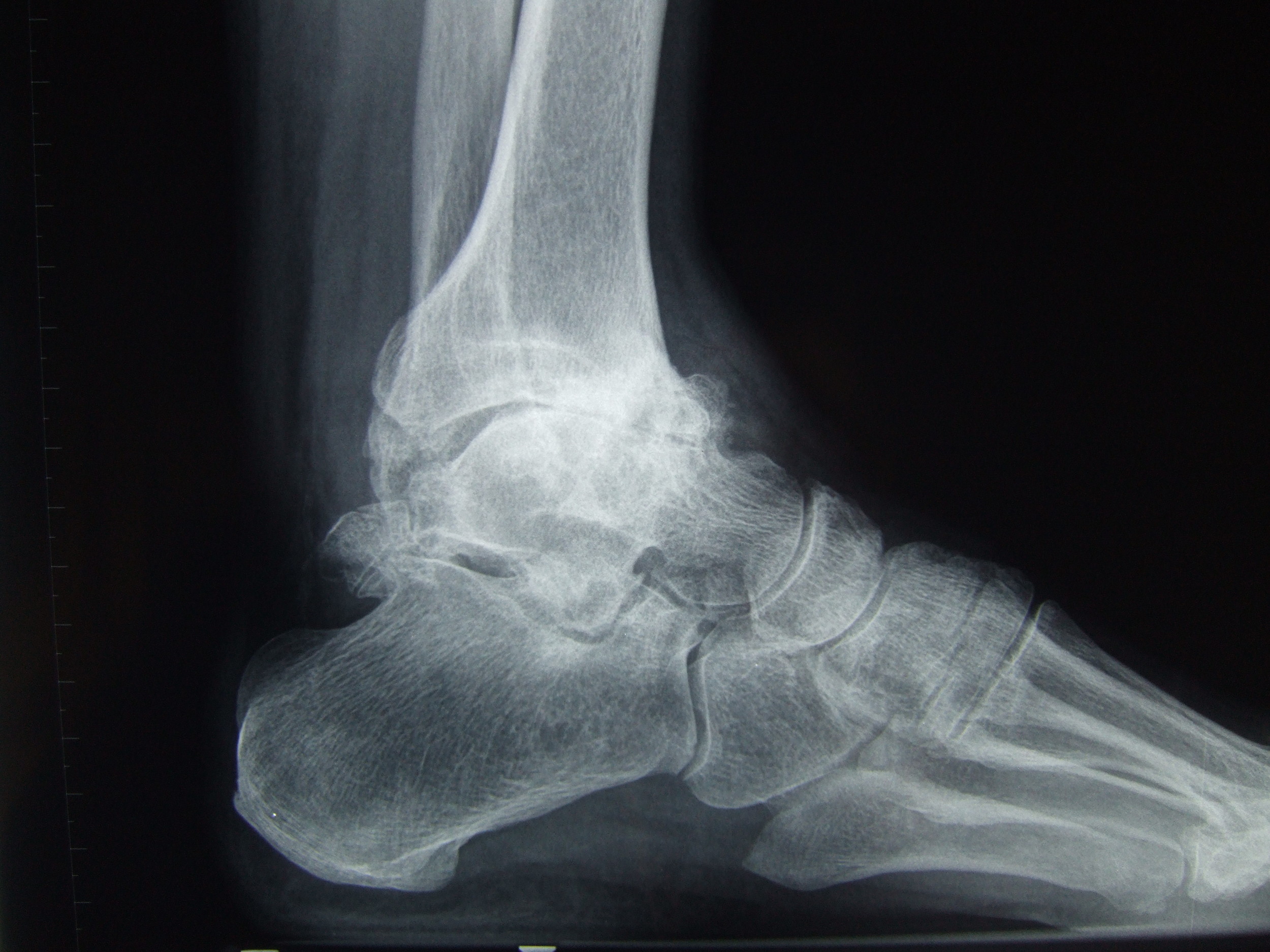
X-ray showing severe arthritis of the ankle joint. Note that there is almost no joint space compared to the normal ankle below (side)
Inflammatory arthritis such as rheumatoid arthritis and gout can damage the joint surface of the ankle as well as other joints in the foot and elsewhere. .
Infections within the joint are fortunately rare, but are another cause of premature arthritis in the ankle.
How is ankle arthritis diagnosed?
If you have ongoing ankle pain you should seek medical advice. Your orthopaedic surgeon will examine your ankle and perform x-rays, these x-rays should be performed with you standing. The x-rays help to confirm arthritis in the ankle but also to identify arthritis in the triple joints below the ankle.
What are the symptoms
Roughening of the joint surface leads to stiffness and pain. Inflammation can cause swelling of the ankle joint. During the early stages, you may have discomfort during exercise which settles with rest. As the arthritis advances, standing or walking becomes painful and eventually pain is experienced even at rest. Occasionally in severe arthritis the joint surface can eroded which may lead to a deformity of the foot with the heel turning in (varus) or out (valgus) when standing.
What is the treatment
Non –operative treatment: There are a number of simple measures that may help control pain.
Pain and inflammation can be controlled with analgesics and anti-inflammatory tablets which can be prescribed by your doctor.
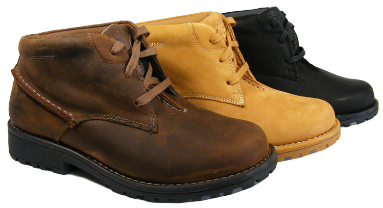
Wearing laced shoes or boots will provide support and limit excessive movement in the ankle.
A number of orthoses or adaptations can help relieve pain, an ankle-foot orthosis is a split that stabilizes the ankle in a right angled posture, it limits movement in the ankle. A ‘Rocker sole’ is an adaptation to the sole of the shoe which makes it curved, this allows you, during walking, to roll over the rocker rather than moving the ankle.
Injections into the ankle are primarily performed to ‘diagnose’ arthritis in the ankle, but sometimes can provide an intermediate period of pain relief.
Ankle Arthrodesis:
An ankle fusion or arthrodesis is an operation which fixes the ankle joint solidly, the body is ‘tricked’ into treating the joint as it would a broken bone and the bones amalgamate into one. The joint surface is removed, the joint is place in its best functional position and solidly fixed with screws. The stiff painful joint becomes a stiff pain-free ankle joint. Despite the ankle joint being fixed, surprisingly almost 50% of the up and down movement of the foot is preserved, this occurs through the adjacent hindfoot joints. The preservation of this up and down movement allows people to walk apparently quite normally, although
you might limp when walking quickly.
Once the joint is fused it stays fused.

The incision for an open ankle fusion or Ankle replacement marked off before surgery and x-rays after a fusion.

X-rays of ankle after ankle fusion with 2 screws across the ankle (side)
Ankle Arthritis
What is ankle arthritis?
The ankle joint is formed by the leg bones (tibia and fibula) and the ankle bone (talus). This joint allows the foot to move up and down. The sideways (inversion/eversion) movement of the heel mainly occurs in the 3 joints (triple joints) under the talus (sub-talar, talo navicular and calcaneocuboid joints). Normally the bones of the ankle joint are covered by smooth articular cartilage which allows the bones to glide over each other as the joint moves freely.
Osteoarthritis may occur spontaneously in the ankle joint but is more commonly associated with previous injury (usually years before). Severe sprains or repeated sprains can damage the articular cartilage of the ankle and lead to progressive arthritis as can fractures around the ankle joint. Click here for link to ankle sprains
An injection of local anaesthetic and steroid into the ankle is often arranged, this will help localize the pain to your ankle.
Occasionally a CT or MRI scan will be needed if arthritis of adjacent joints is a concern.

Well preserved ankle

Operative treatments: Early arthritis may involve only localized areas of the joint surface which may be amenable to treatment by key hole surgery using ankle arthroscopy as a day-case.
When the pain becomes severe and affects your quality of life, interferes with your usual daily activities or causes you to regularly lose sleep despite non operative treatments, then an surgery is indicated.
If there is progressive deformity of the ankle joint then surgery may be recommended before damage occurs to the other joints adjacent to the ankle (eg sub-talar joint).
The two surgical treatments commonly used to treat severe ankle arthritis are ankle fusion or ankle replacement.
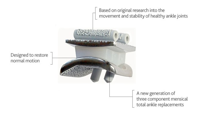
Total Ankle Replacement:
A total ankle replacement is an operation that removes bone and cartilage from the arthritic ankle joint surface and replaces them with metal surfaces and a polyethylene spacer The metallic implants are lined on their deep surface with a roughened layer that stimulates bone ingrowth and a joint surface that is highly polished. An ankle replacement preserves a degree of ankle movement (usually not full movement).
Preservation of ankle movement is especially valuable in those patients who have arthritis and stiffness affecting other joints in the foot, which might be the case in conditions such as rheumatoid arthritis. In these patients an ankle fusion would lead to a lot of stiffness due to lack of compensatory movements in the adjacent hindfoot joints.
The results of ankle replacement have improved over the last ten years but to date the new generation replacements haven’t achieved results that compare to hip and knee replacements.
Over time joint replacements will wear out or fail, as is the case with any machine. When an ankle replacement wears out there is frequently a loss of bone above and below the metal implants. As the ankle bone (talus) is a relatively small bone, there is often insufficient bone to allow a new implant to be attached or to allow an ankle fusion in which case a fusion of the ankle to the heel (subtalar joint) is required which produces more stiffness than an isolated ankle fusion.
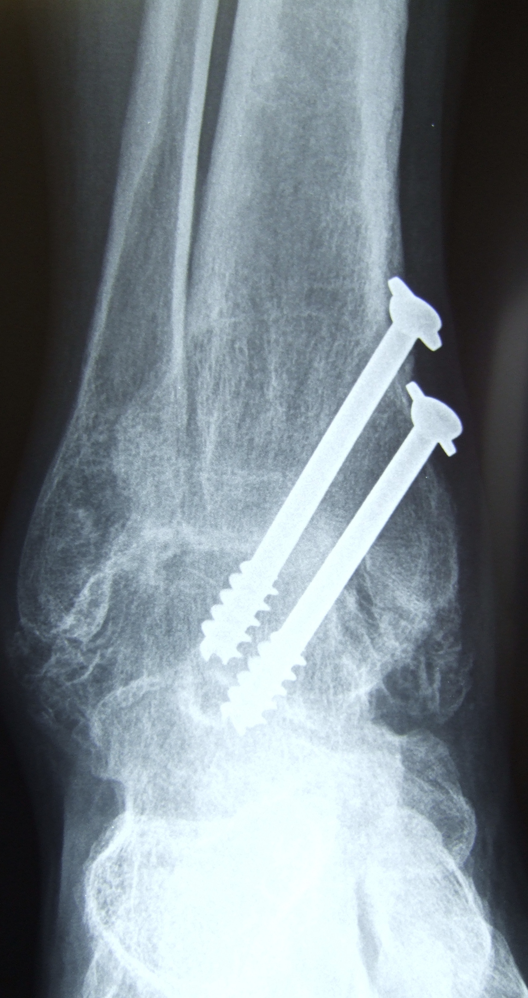
X-rays of ankle after ankle fusion with 2 screws across the ankle (front)

A fusion of the ankle and subtalar (joint below ankle) following a failed total ankle replacement.
Be sociable..share!